
Recorded Webinars
Lung Ultrasound for COVID-19
This course will outline a 4-step process for evaluating valve function.
Step 1: 2D evaluation of the valve (eyeball method)
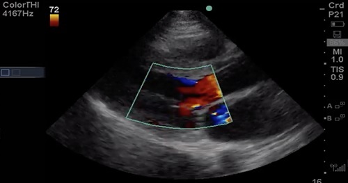
Step 2: Assess the valve with color Doppler for Mitral Regurgitation (MR) or Aortic Regurgitation (AR)
Step 3: Assign relative importance of lesion
This course will compare Transthoracic Echocardiography (TTE) and Transesophageal Echocardiography (TEE), outline clinical questions with point-of-care TEE, describe how to get started with TEE (politics, cost, logistics), discuss safety and training, review a suggested TEE protocol, and give several case examples using TEE.
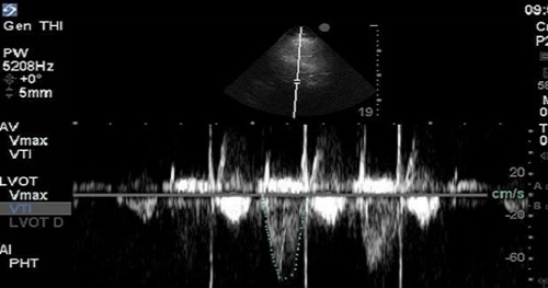
Determination of Stroke Volume (SV) is perhaps the most essential of all the “advanced” techniques for point-of-care echocardiography. It’s considered advanced, because it relies on quantitative spectral Doppler techniques, which are not routinely part of the core point-of-care echocardiography curricula offered for most specialties at the time of this publication. This technique is the most practical and intuitive gateway to hemodynamic understanding of echocardiography and is a powerful adjunct in the assessment of Left Ventricular (LV) function.
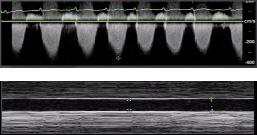
There are a number of different data points that can be collected that inform us of the function and loading conditions of the Right Ventricle (RV): Shape and size of the RV, Inferior Vena Cava (IVC), Tricuspid Angular Plane Systolic Excursion (TAPSE), and Right Ventricular Systolic Pressure (RVSP).
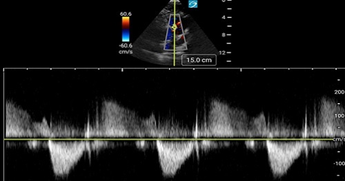
Doppler echocardiography is the language of flow in and around the heart. In order to evaluate hemodynamics in and around cardiac valves, cardiac pressures or in the calculation of Stroke Volume (SV), one must speak the language of Doppler.
This introduction to Doppler principles and how they relate to point-of-care echocardiography will frame the knowledge you need to engage some particularly valuable hemodynamic techniques!