Enrico Storti

Anesthesia and ICU Director
Unit Coordinator of the Emergency Department
Maggiore Hospital
Lodi, Italy
Family name
Storti
Unit Coordinator of the Emergency Department
Maggiore Hospital
Lodi, Italy
Saint Mary’s Hospital, an acute care community teaching hospital in Waterbury, Connecticut demonstrated the clinical and financial benefits of an ERAS program that includes ultrasound-guided regional anesthesia (USGRA). Since the October 2015 launch of the opioid-sparing ERAS program, Saint Mary’s has seen striking improvements in the safety and quality of care for patients undergoing colorectal surgery, along with substantial reductions in opioid usage.
St. Joseph’s has the second-busiest Emergency Department in the United States, seeing 175,000 patients in 2016. St. Joseph’s Emergency Department launched the ALTOSM (ALTernatives to Opioids) Program in January 2016.
The program uses targeted non-opioid medications, trigger point injections, nitrous oxide, ultrasound-guided nerve blocks, and even meditation, to help ease pain in patients with acute injuries like broken bones, and ongoing issues like migraine headaches.
This course will outline a 4-step process for evaluating valve function.
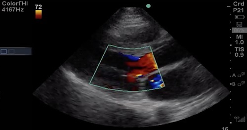
Step 1: 2D evaluation of the valve (eyeball method)
Step 2: Assess the valve with color Doppler for Mitral Regurgitation (MR) or Aortic Regurgitation (AR)
Step 3: Assign relative importance of lesion
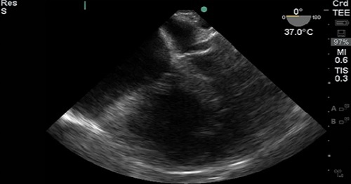
This course will compare Transthoracic Echocardiography (TTE) and Transesophageal Echocardiography (TEE), outline clinical questions with point-of-care TEE, describe how to get started with TEE (politics, cost, logistics), discuss safety and training, review a suggested TEE protocol, and give several case examples using TEE.
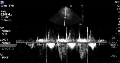
Determination of Stroke Volume (SV) is perhaps the most essential of all the “advanced” techniques for point-of-care echocardiography. It’s considered advanced, because it relies on quantitative spectral Doppler techniques, which are not routinely part of the core point-of-care echocardiography curricula offered for most specialties at the time of this publication. This technique is the most practical and intuitive gateway to hemodynamic understanding of echocardiography and is a powerful adjunct in the assessment of Left Ventricular (LV) function.
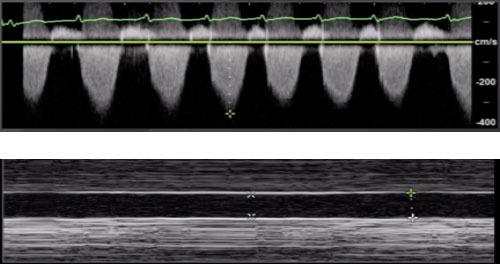
There are a number of different data points that can be collected that inform us of the function and loading conditions of the Right Ventricle (RV): Shape and size of the RV, Inferior Vena Cava (IVC), Tricuspid Angular Plane Systolic Excursion (TAPSE), and Right Ventricular Systolic Pressure (RVSP).