Recorded Webinars
Webinar: POCUS and Undifferentiated Shock

Cameron Baston, MD, MSCE, is a clinician advisor for Penn Health-Tech and an Assistant Professor of clinical medicine in the Department of Medicine at the University of Pennsylvania's Perelman School of Medicine. He serves as associate program director for the Pulmonary and Critical Care Medicine fellowship, and as director of clinician-performed ultrasound for the Department of Medicine. He has an interest in helping create low-cost medical devices in the resource-limited Critical Care setting and works with several organizations on POCUS and Critical Care education. A mechanical engineer, epidemiologist, Critical Care physician, and medical educator, he spends about 2/3 of his time caring for critically ill patients, and the remainder working on education innovation and health technology.
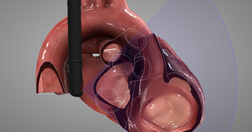
Point of care Transesophageal Ultrasound (TEU) is an emerging modality that offers bedside clinicians the ability to assess critically ill patients quickly. A Transesophageal Echocardiogram, or TEE, is an alternative way to perform an echocardiogram. A specialized transducer containing an ultrasound transducer at its tip is passed into the patient's esophagus. This allows image and Doppler evaluation which can be recorded.