Cardiac Imaging 2 - Valvular Assessment (English Only)
Temps du cours: 1h 24min
This course will outline a 4-step process for evaluating valve function.
Step 1: 2D evaluation of the valve (eyeball method)
- Morphology, mobility, vegetations
Step 2: Assess the valve with color Doppler for Mitral Regurgitation (MR) or Aortic Regurgitation (AR)
- Classify as severe or non-severe
- If severe, escalate to an advanced echo
- If non-severe, classify importance and possibly escalate to an advanced echo
Step 3: Assign relative importance of lesion
- Devastating, important, or incidental
Step 4: Consider appropriate clinical action (acute interventions, referral, advanced echocardiogram etc)
of 4 sections terminées
Cardiac 2 - Valvular Assessment: Introduction
Introductory material and disclaimer
Cardiac 2 - Valvular Assessment: Lesson
Bedside Valvular Assessment
Cardiac 2 - Valvular Assessment: Post-test
Assess your knowledge of the material presented in the lesson with a post-test. Score 80% or higher to receive a certificate of completion.
Cardiac 2 - Valvular Assessment: Additional Resources
View additional material related to this course
Description
- Mitral Valve
- Aortic Valve
Description
- Tricuspid Valve
- Mitral Valve
Description
- Mitral Valve
- Aortic Valve
Description
- Aortic Valve
- Mitral Valve
Description
- Mitral Valve
Description
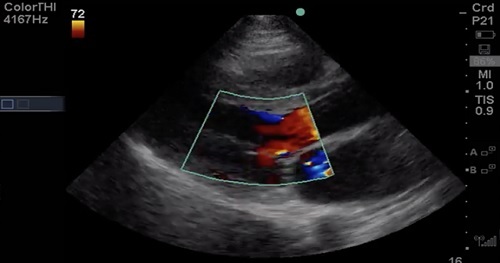
- Vena Contracta through the aortic valve in a zoomed in image from a Parasternal Long Axis (PLAX) view
Description
- Vena Contracta through the aortic valve in a zoomed in image from a Parasternal Long Axis (PLAX) view
Description
- Vena Contracta of Mitral Regurgitant (MR) Jet
- Left Ventricle (LV)
- Right Ventricle (RV)
- Right Atrium (RA)
Description
- Tricuspid Valve
- Mitral Valve
- Septum


